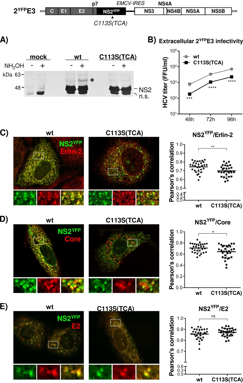
FIG 6.
Impaired NS2 palmitoylation reduced the colocalization of NS2YFP with Erlin-2 and Core but not with E2. (Top) Schematic of 2YFPE3, which has an organization identical to that of 2E3 but with NS2YFP replacing NS2. (A) Western blot detection of palmitoylated NS2 in cell lysates derived from wt or C113S(TCA) mutated 2YFPE3 RNA replicating cells following APEGS assay. The PEGylated NS2YFP is indicated by an asterisk. n.s., nonspecific bands. (B) Extracellular HCV titer determined using culture supernatants collected at different time points postelectroporation of wt or NS2YFP/C113S(TCA) mutated 2YFPE3 RNA. (C to E) Confocal imaging (left) and Pearson’s correlation coefficient analysis (right) of 30 different images to detect the colocalization of NS2YFP with Erlin-2 (C), Core (D), and E2 (E) in cells at day 3 postelectroporation of wt or NS2YFP/C113S(TCA) mutated 2YFPE3 RNA. The asterisks indicate statistically significant differences between paired values based on an unpaired Student t test with Welch’s correction. *, P < 0.05; ** P < 0.005. A difference with a P value of >0.05 was considered not significant (ns).

