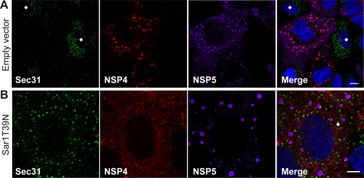
FIG 5.
Rotavirus infection disrupts COPII vesicle protein localization at ER exit sites and expression of Sar1T39N disrupts NSP4 trafficking to viroplasms. (A and B) Representative confocal images of rotavirus-infected cells transfected with empty plasmid (A) or Sar1T39N-expressing plasmid (B). Cells were probed with rabbit peptide-specific antibody αNSP4114–135, guinea pig antibody αNSP5 to detect viroplasms, and monoclonal antibody αSec31 to identify ER exit sites. Noninfected cells are indicated by an asterisk in panel A. Scale bars, 5 μm.

