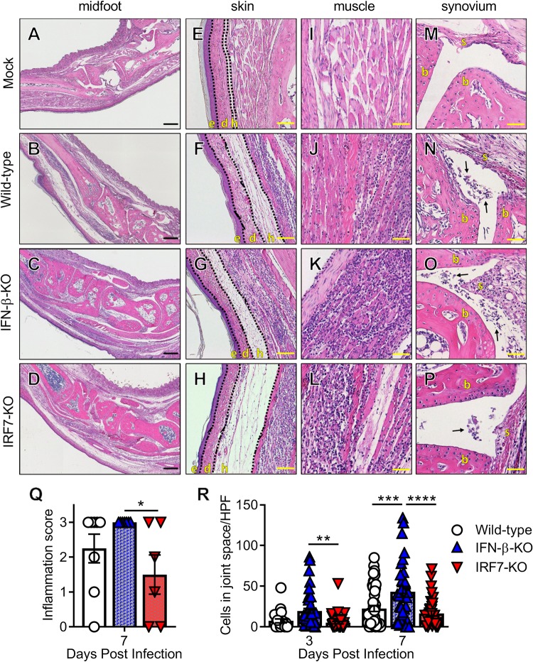
FIG 4.
IFN-β-KO but not IRF7-KO mice have increased immune cell infiltration in the joints during acute CHIKV infection. Wild-type, IFN-β-KO, or IRF7-KO mice were mock treated or inoculated with 103 PFU of CHIKV, and ipsilateral ankles/feet were processed at 3 or 7 dpi for histological analysis by hematoxylin and eosin staining. (A to P) Representative images of sections of the midfoot (tiled), skin, muscle, and synovium at 7 dpi. (E to H) The skin and associated tissue sections show the epidermis (e), dermis (d), and hypodermis (h). (M to P) The synovium sections show synovium (s) and bone (b), with arrows indicating immune infiltrates into the synovial cavity. Bars = 200 μm (A to D), 50 μm (E to H and M to P), and 100 μm (I to L). (Q) Feet and ankles were scored for histological damage as described in Materials and Methods. (R) Quantification of inflammatory cells per high-power field (HPF) in the midfoot joint spaces as described in Materials and Methods. For panels Q and R, data are pooled from at least two experiments with 4 to 6 mice per group. Data were analyzed using one-way ANOVA (Q) or two-way ANOVA (R) with Tukey’s posttest. All data are presented as means ± SEM. (*, P < 0.05; **, P < 0.01; ***, P < 0.001; ****, P < 0.0001).

