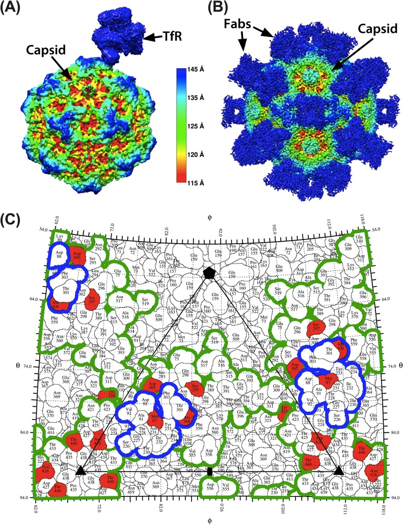
FIG 2.
Structural context of natural CPV variants and their relation to TfR and MAb Fab footprints. (A and B) High-resolution structure of the transferrin receptor (A) and monoclonal antibody Fabs (B) bound to the CPV capsid colored by capsid radial distance identifies the sites of contact. (C) One asymmetric unit of CPV capsid, showing the footprint of the TfR (blue outline) and the outline of the combined footprints of eight different antibodies (green outline). Red residues varied during evolution of CPV in dogs. All structures were determined by cryoEM. See main text for references.

