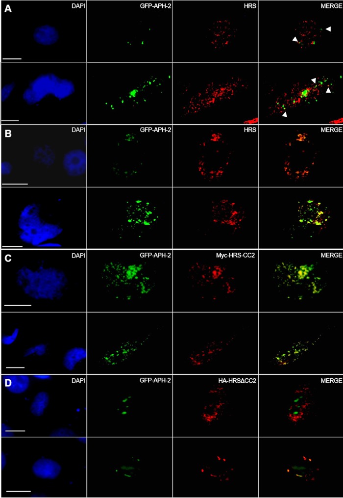
FIG 3.
HRS and APH-2 colocalize in endocytic structures in a CC2-dependent manner. HeLa cells were transfected with 750 ng of GFP-APH-2 (green) and 250 ng of control plasmid (A), HA-HRS (B), Myc-HRS-CC2 (C), or HA-HRSΔCC2 (D) expression plasmids; 24 h posttransfection, endogenous HRS and exogenous HRS were detected using anti-HRS (A, B, and D) or anti-Myc antibodies (C), followed by Alexa Fluor 594-conjugated secondary antibody (red). Nuclei were stained with DAPI (blue). Two representative images are shown per condition. The arrowheads indicate colocalization of HRS and GFP-APH-2. Scale bars, 10 μm.

