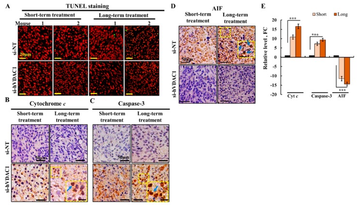
Figure 4.
si-hVDAC1-TTs showed no apoptotic cell death, despite changes in the expression levels of pro-apoptotic proteins. (A) Paraffin-embedded sections from hVDAC1-TTs or si-NT-TTs following short- or long-term treatment were subjected to TUNEL staining, as described previously [1]. (B) Representative sections from si-NT-TTs and si-hVDAC1-TTs IHC-stained for Cyto c (B), caspase 3 (C), and apoptosis-inducing factor (AIF) (D), using specific antibodies, and quantitation of the staining intensity (E) using a panoramic microscope and HistoQuant software (Quant Center 2.0 software,1.15.1 RTM 3DHISTECH Ltd) are shown as fold change (FC) relative to the levels in si-NT-TTs Inset shows nuclear localization, marked by arrows, bar = 20 μm. Data are expressed as mean ± SEM, (n = 3 tumors), (*** p < 0.001).

