Figure 6.
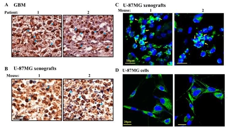
TSPO expression levels decreased in si-hVDAC1 treated U-87MG-derived tumors. Representative sections from si-NT-TTs and si-hVDAC1-TTs, treated for short or long periods were immunohistochemically stained for TSPO (A) and the staining intensity was quantified using a panoramic microscope and analyzed by HistoQuant software (Quant Center 2.0 software, 3DHISTECH Ltd) (B) are shown as the intensity in relative units (RU). Data are expressed as mean ± SEM, (n = 3 tumors). Confocal fluorescence microscopic images of immuno-stained sections from si-NT-TTs and si-hVDAC1-TTs, treated for short (C) or long (D) periods stained with anti-TSPO or anti-VDAC1 antibodies. Nuclei were stained by DAPI. Enlargement of the corresponding squad area is presented with arrows indicating the nuclear localization of TSPO.

