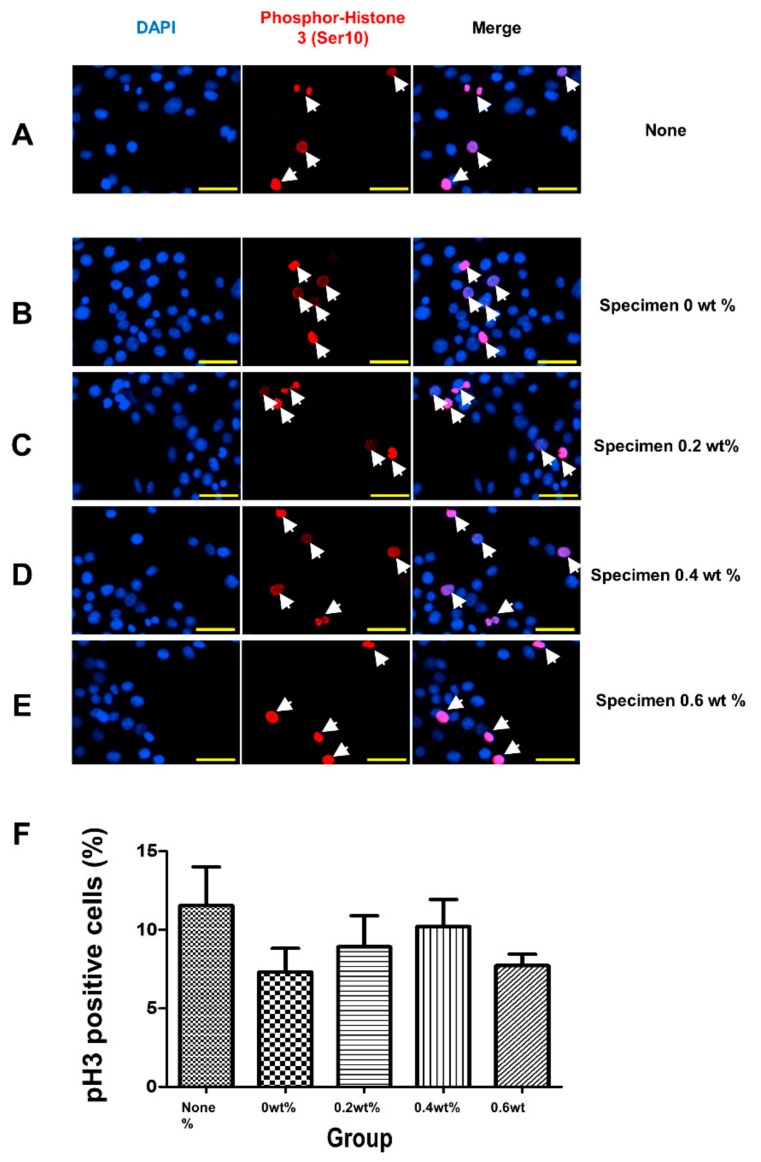
Figure 7.
Analysis of mitotic capacity after coating material administration. (A–F) Immunofluorescence analysis of phospho-Histone 3 (Ser 10) proteins in non-treated (A) or coating material-challenged (B–E) cells. The concentrations of materials were indicated on the panel. Cells were incubated with specimens for 24 h. Nuclear 4′,6-diamidino-2-phenylindole staining is shown in blue. (F) The quantification of phospho-Histone 3 (Ser 10)-positive cell proportions was analyzed. Nuclear 4’,6-diamidino-2-phenylindole (DAPI) staining is shown in blue. The scale bar is 50 µm.

