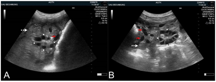
Figure 6.
Transabdominal ultrasonographic images of ovaries in sows in estrus with round-oval (A) and polygonal pre-ovulatory follicles (B). (A,B) Squares mark the transversal and longitudinal dimension of the ovaries. Example cross-sections of the uterine horns (white, dotted arrows) and blood vessels (red arrows) are identified. Scale bars on right margins in 0.5 cm steps.

