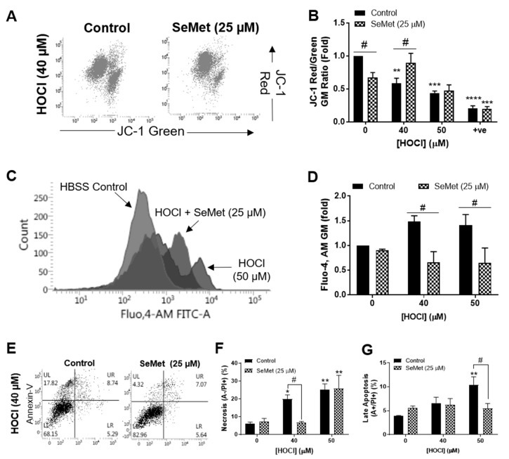
Figure 1.
Selenomethionine (SeMet) supplementation protects against H9c2 cellular dysfunction elicited by HOCl. H9c2 cells (1 × 10⁵ cells mL−1) serum-starved and supplemented with (hatched bars) or without (solid bars) SeMet (25 µM) for 24 h were exposed to HOCl for 1 h with or without SeMet (25 µM) and analysed immediately for ΔΨm and intracellular Ca2+ accumulation using JC-1 and the fluorescent Ca2+ indicator Fluo,4-AM, respectively and the extent of cell death measured using Annexin-V/propidium iodide (PI) staining. (A) Representative dot plots of JC-1 staining with (B) quantification of ΔΨm immediately following exposure to HOCl and SeMet supplementation or CCCP (0.1 mM) as a positive control (+ve). (C) Representative histogram flow plot of Fluo,4-AM staining with (D) quantification of intracellular Ca2+ accumulation immediately following exposure to HOCl and SeMet supplementation. (E) Representative dot plots of Annexin-V/PI staining with (F) quantification of necrotic cells and (G) late apoptotic cells immediately following exposure to HOCl and SeMet supplementation. Data are expressed as a percentage of the whole cell populations as mean ± S.E.M. from n = 3 biological repeats performed in triplicate. (B,D) Data are expressed as a fold change over respective controls assayed in the absence of oxidant and SeMet as mean ± S.E.M. from n = 3 biological repeats performed in triplicate. * p < 0.05, ** p < 0.01, *** p < 0.001, **** p < 0.0001 vs. Hank’s buffered salt solution (HBSS) control (0 µM), # p < 0.05 vs. control cells assayed in the absence of SeMet (0 µM) as determined by two-way ANOVA with Bonferroni post-hoc testing.

