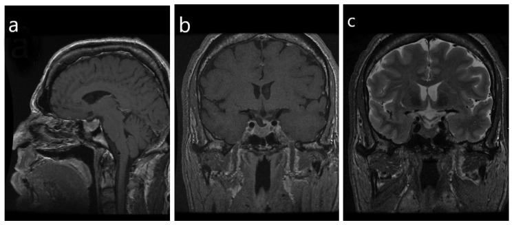
Figure 2.
Radiologic imaging of a densely-granulated somatotroph tumor. (a) Note the frontal bossing, prognathism and thickening of the scalp with a “rug sign” that are evident on the sagittal view; (b) in the coronal view the sella is enlarged, with evidence of a macrotumor. The infundibulum is deviated to the left, with an associated focal defect versus a depression in the sellar floor and an inferior herniation of pituitary glandular tissue. There is ptosis of the optic chiasm, with no evidence of mass effect on the chiasm, (c) The tumor does not have a high T2 signal, however, a hypointensity signal is noted within the optic chiasm and proximal cisternal segments of the bilateral optic nerves.

