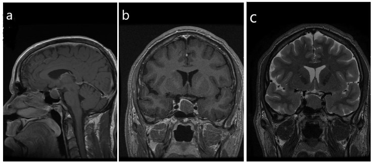
Figure 4.
Radiologic imaging of a sparsely-granulated somatotroph tumor. (a,b) There is a homogeneous hypoenhancing macrotumor that measures 1.5 × 1.7 × 1.9 cm, with mild suprasellar extension and mild mass effect on the optic chiasm. There is mild abutment of the left cavernous ICA, consistent with Knosp grade 1; Note the lack of features of florid acromegaly compared with Figure 1. (c) T2-weighted imaging, coronal view reveals that the tumor is homogeneous and isointense to the gray matter.

