Figure 4.
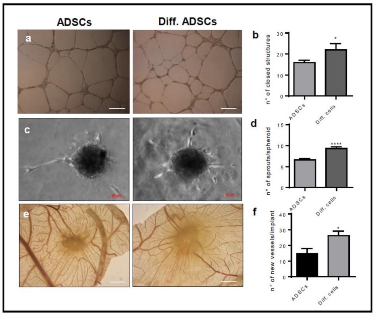
The differentiation into ADSC derived beige cells positively modulates the pro-angiogenic ability of ADSCs. (a) Confluent monolayers of ADSCs and ADSC derived beige cells (Diff.cells) were cultured in 24 wells. Two-hundred microliters of growth factor reduced Cultrex were added on the monolayers, and HUVEC cells were plated on gel. After 18 h, the formation of capillary-like structures was examined using a Zeiss Axiovert 200 M microscope (Scale bar 500 μm). Data are the mean ± SEM of three measurements per sample. * p < 0.05, Student’s t-test. (b) The number of closed structures. (c–d) HUVEC spheroids embedded in collagen gel and plated on an ADSC monolayer. The formation of radially growing sprouts was evaluated after 24 h of incubation. Data are the mean ± SEM of three independent experiments (* p < 0.05, **** p < 0.001, one-way ANOVA followed by Bonferroni’s test versus the control). (e–f) Alginate beads containing 3 × 10⁴ ADSCs or ADSC derived beige cells were implanted on the top of chick embryo chorioallantoic membranes (CAMs) at Day 11 of development. After 72 h, newly formed blood vessels converging toward the implant were counted in ovo at 5× magnification using an STEMI SR stereomicroscope equipped with an objective f equal to 100 mm with adapter ring 475,070 (Carl Zeiss) (Scale bar 2 mm). Data are the mean ± SEM (n = 6–8) (* p < 0.05, one-way ANOVA followed by Bonferroni’s test versus the control).

