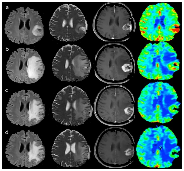
Figure 3.
Pseudoprogression during immunotherapy of a LowNK and hypermethylated case - Left to right: FLAIR, ADC map, T1-enhanced and CBV map. (a) Jan 2015, MRI-pre, after surgery and radio-chemotherapy (steroid dose 2 mg Dexamethazone, clinical condition stable): GBM showing contrast enhancement (T1) and hyper-perfusion with high CBV (red-colored), slight edema (FLAIR) and non-homogeneous ADC restriction as in hypercellularity; (b) Mar 2015, MRI-2 mo during immunotherapy (no steroid therapy, clinical condition stable): enlargement of enhancing volume and of edema with mass effect and reduction of hyper-perfusional intensity (yellow to green CBV with small red spots) and persistent ADC un-homogeneity; (c,d) Jun and Aug 2015, MRI-4 and -6 mo after immunotherapy (no steroid therapy clinical condition stable): reduction of enhancing volume, edema and CBV intensity (green) and volume of hyper-perfusion with less restricted ADC.

