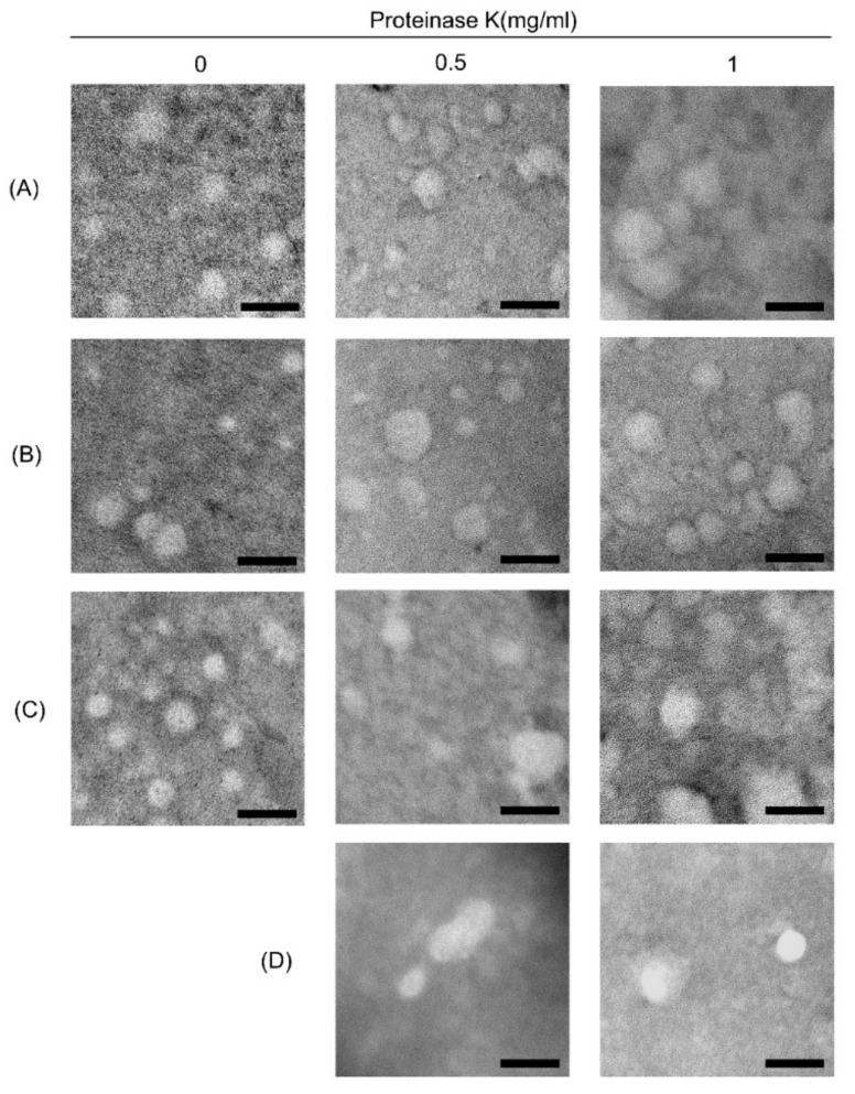
Figure 3.
Electron microscopic image of exosomes following different isolation method. ELVs were purified using (A) SBI, (B) LT, (C,D) QIAGEN treated with different concentration of proteinase K (0, 0.5, 1mg/mL), and (D) additionally treated with HCl for acidification of samples to approximately pH 4.0. Diluted suspension containing ELVs was placed on formvar-carbon coated grid and negatively stained with uranyl acetate. Round-shaped vesicles with a heterogeneous size from 30 to 150 nm diameter were clearly visualized in all preparations. The horizontal bars indicate 100 nm.

