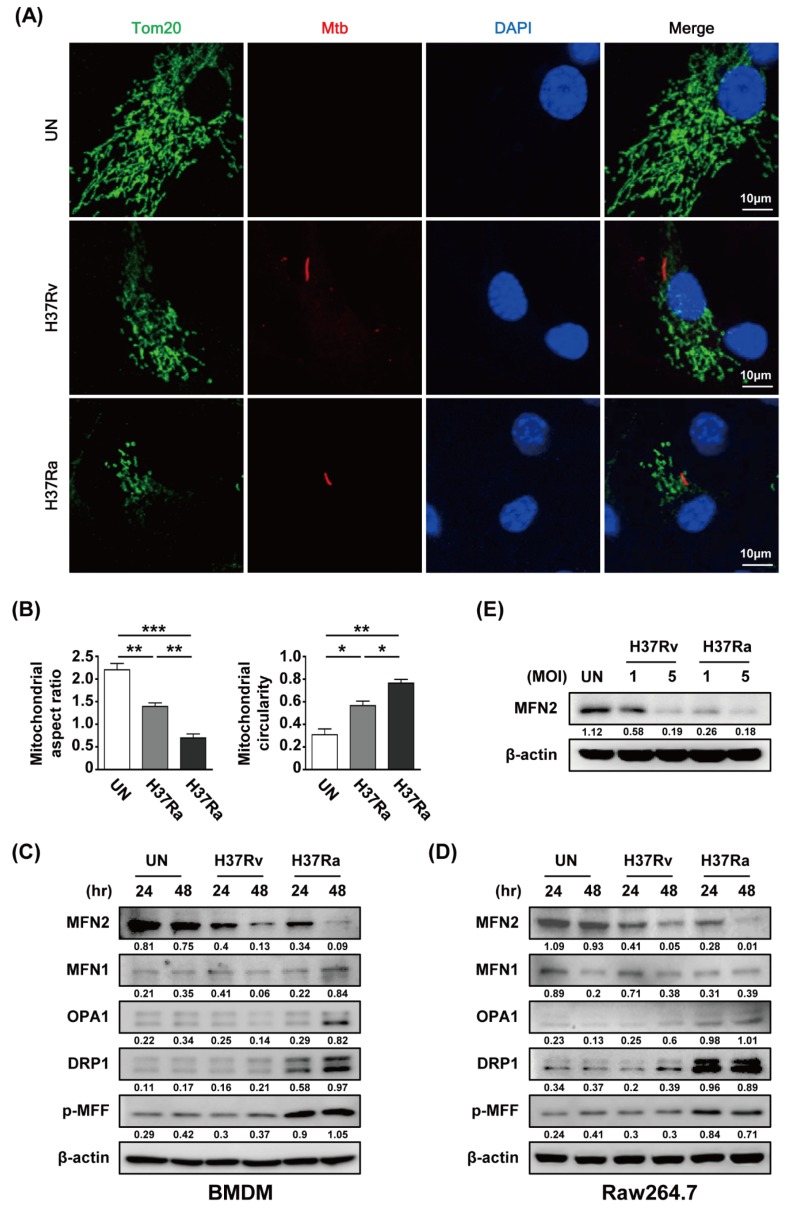
Figure 2.
Mtb induces mitochondrial fragmentation in macrophages. (A) BMDMs were infected with RFP-labeled Rv or Ra (red), incubated for 48 h, stained with Tom20 to detect mitochondria (green) and DAPI to visualize nuclei (blue), and visualized using confocal microscopy. (B) Quantification of the mitochondrial morphology (aspect ratio; left, and circularity; right) in (A). (C,D) Cells were harvested and subjected to western blotting for mitochondrial fusion/fission factors (such as MFN1/2, OPA1, Drp1, and p-MFF); β-actin was used as the loading control. (E) BMDMs were infected with Rv or Ra (MOI = 1 to 5) for 3 h, and then incubated 48 h. Western blotting analysis was performed using antibodies directed against MFN2 and β-actin. Data are representative of three independent experiments. Statistically significant differences are indicated; * p < 0.05, ** p < 0.01, and *** p < 0.001.

