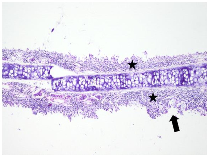
Figure 6.
Light micrographs of the rat’s nasal septum, astaxanthin group, at 14 days after the intranasal intervention. Diffusely, the lamina propria of the mucosa covering the nasal septum is markedly expanded by many macrophages admixed with lymphocytes and few plasma cells (black star). The covering mucosa is focally erosive and occasionally hyperplastic (arrow); H&E, ob ×10.

