Figure 2.
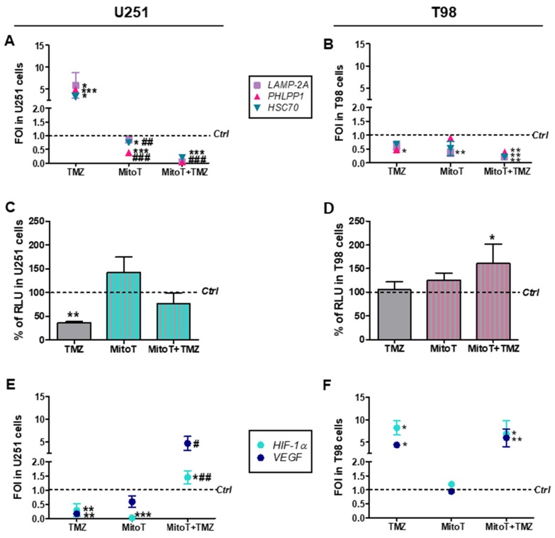
Involvement of chaperone-mediated-autophagy (CMA) after Temozolomide and MitoTempo (MitoT) treatment. (A) Gene expression analysis for CMA-related genes (LAMP2A, HSC70, PHLPP1) analyzed by means of Real-time PCR in U251 and (B) T98 cells after treatment with 100 µM Temozolomide (TMZ) ± 25 µM MitoT. Data were normalized for β-ACTIN, and the ΔΔCt values were expressed as FOI of the ratio between treated and control cells. * p < 0.05; ** p < 0.01; *** p < 0.001 treated vs. control cells. # p < 0.05, ## p < 0.01, ### p < 0.001 vs. TMZ-treated cells. (C) Biochemical assay for HIF-1α activity in U251 and (D) T98 cell lines. Data were expressed as RLU, obtained normalizing luciferase counts for the amount of proteins quantified by Bradford assay. ** p < 0.01 vs. control cells. (E) Gene expression analysis for HIF-1α and vascular endothelial growth factor (VEGF) expression in U251 and (F) T98 cells. * p < 0.05; ** p < 0.01 vs. control cells; # p < 0.05, ## p < 0.01 vs. TMZ-treated cells. Mean values ± SD of three independent experiments.

