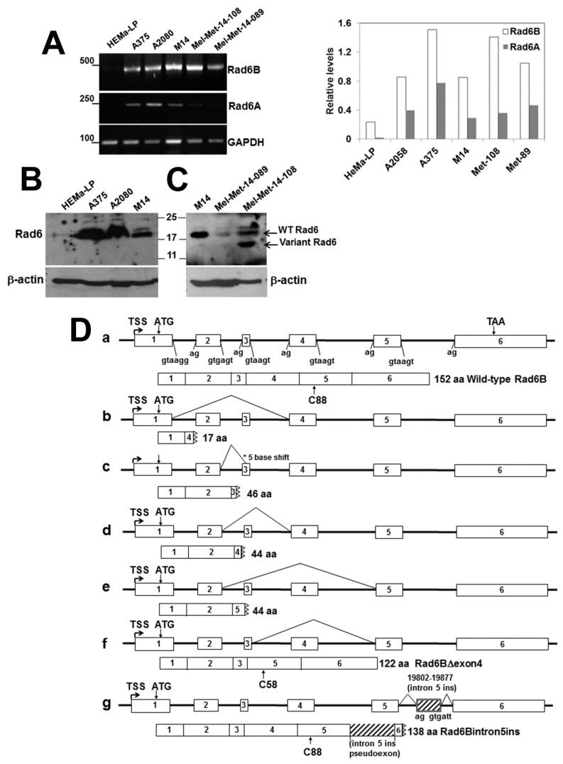
Figure 2.
RAD6B is robustly expressed in melanoma lines and shows abnormal processing of transcript splicing. (A) RT-PCR analysis of RAD6A and RAD6B in normal (HeMa-LP) melanocytes, melanoma cell lines and clinical melanoma brain metastasis samples Mel-Met-14-108 and Mel-Met-14-089. RAD6A and RAD6B levels normalized to GAPDH are shown in the graph on the right. (B) and (C), Western blot analysis of RAD6 in HeMa-LP, melanoma lines and clinical melanomas. Note that since the RAD6 antibody does not distinguish RAD6A and RAD6B, the protein is indicated as RAD6. The presence of a 14 kDa variant RAD6 protein in clinical melanoma is indicated. (D) (a) Schematic structure of the human RAD6B gene and splice junctions. The wild-type protein with the active cysteine (C)88 is indicated below. (b–g) Schematic mutations of RAD6B splicing mutations identified in melanoma lines and clinical melanoma brain metastases. The 122 amino acid RAD6BΔexon4 mutant (f) was identified in M14 and A2058 melanoma lines, and the 138 amino acid RAD6Bintron5ins (g) splice mutant was identified in the M14 line. The positions of the active cysteine residue in the splice mutants are shown.

