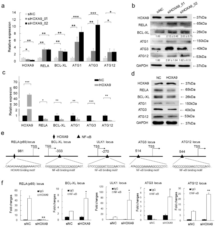
Figure 2.
HOXA9 represses the expression of NF-κB and its downstream apoptotic and autophagic genes by directly binding to the promoter region of NF-κB. (a–d) The mRNA or protein expression levels of HOXA9, RELA (p65), BCL-XL, ULK1, ATG3, and ATG12 were detected by qRT-PCR (n = 3) or western blot in cSCC cells after knockdown of HOXA9 by two siRNAs or overexpression of HOXA9. The qRT-PCR data were normalized to GAPDH gene expression. In western blots, GAPDH was used as a loading control. The bands of BCL-XL and ATG12 were densimetrically quantified (n = 3). (e) Predicted binding site of HOXA9 (diamond) at the promoter of RELA (p65) or binding sites of RELA for on the promoter regions of BCL-XL, ULK1, ATG3, and ATG12 by rVista (https://rvista.dcode.org/). (f) The binding enrichment of HOXA9 at the RELA locus or RELA at the loci of BCL-XL, ULK1, ATG3, and ATG12 was detected by ChIP-qPCR after knockdown of HOXA9. One-Way ANOVA and Dunnett’s multiple comparison test. Means ± s.d., * p < 0.05, ** p < 0.01, *** p < 0.001.

