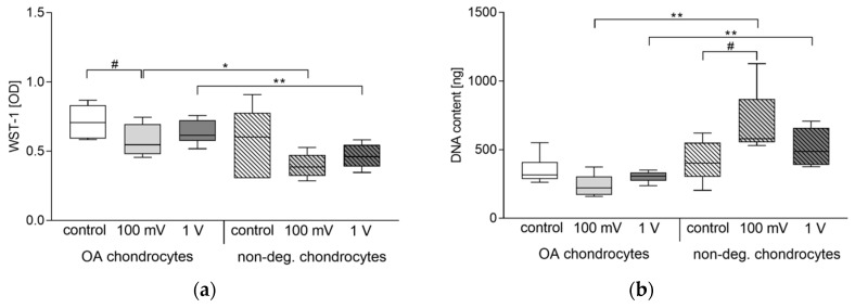
Figure 4.
Cellular activity of human chondrocytes following electrical stimulation with 1 kHz and either 100 mV or 1 V. Chondrocytes derived from non-degenerative (n = 4) or osteoarthritic (OA) cartilage (n = 6) were seeded on collagen scaffolds and stimulated over a period of seven days. Afterwards, metabolic activity was determined via water-soluble tetrazolium salt (WST-1) assay (a) and DNA content was analyzed by peqGOLD Tissue DNA Mini Kit (b). Data are presented as boxplots. Statistical analysis within a stimulation group was performed with Friedman test (# p < 0.05). To compare two samples between OA and non-degenerative chondrocytes, Mann-Whitney-U-test was performed (* p < 0.05, ** p < 0.01).

