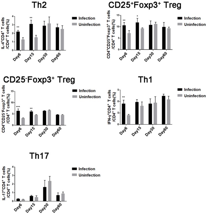
Figure 1.
Differentiation of Treg, Th1, Th2, and Th17 cells in mice during T. spiralis infection: For each of three independent experiments, 16 female BALB/c mice were each infected with 400 T. spiralis ML. Four mice were randomly sacrificed at different stages post-infection (intestinal phase, day 6; newborn larva (NBL) migration, day 15; larva capsule, day 30; and convalescent phase, day 60); Th2, Treg, Th1, and Th17 CD4+ T cells in the spleen were measured by fluorescence-activated cell sorting (FACS). Data are shown as means ± SDs; * p < 0.05; ** p < 0.01; *** p < 0.001.

