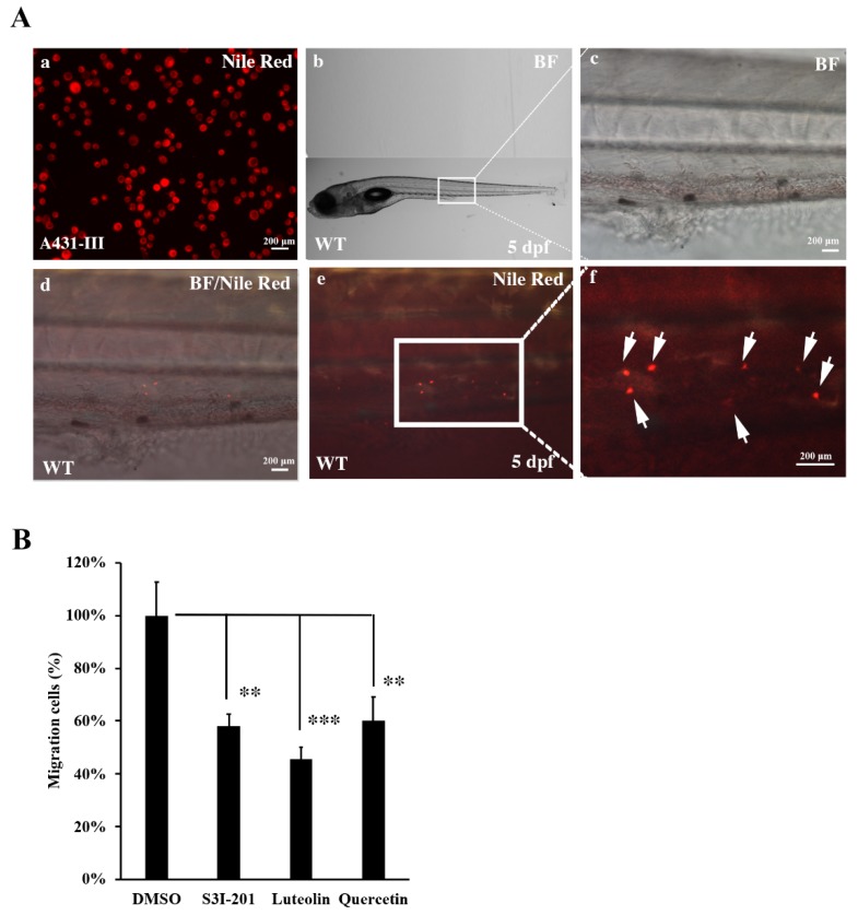
Figure 7.
Metastasis of A431-III cells were reduced by suppression of Src/Stat3 signaling in zebrafish. (A) A431-III cells were stained with Nile red and microinjected into the pericardiac space of zebrafish larvae at 2 days post-fertilization (dpf). (a) A431-III cells were stained with Nile-Red. (b) The bright field view of 5-dpf zebrafish larvae. Bright field view (c), bright field combined with fluorescent view (d) and fluorescent view (e) of migrative tumor cells enlarged from figure b. (f) Fluorescent view of migrative tumor cells (white arrow) enlarged view from figure e. The number of migrative tumor cells were measured by fluorescent microscope. (B) Measurement of metastatic tumor cell numbers pretreated with 0.1% DMSO (DMSO), 400 μM S3I-201 (S3I-201), 20 μM luteolin, and 40 μM quercetin in zebrafish larvae. Statistical significance between groups were analyzed by a one-way ANOVA with Tukey’s test (** p < 0.01; *** p < 0.001). BF: bright field.

