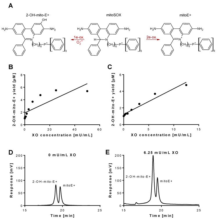
Figure 1.
Detection of superoxide generation by xanthine oxidase by mitoSOX HPLC. (A) Structures of mitoSOX and its oxidation products 2-OH-mito-E+ (left) and mitoE+ (right). (B,C) The yield of the superoxide-specific mitoSOX oxidation product 2-OH-mito-E+ in dependence of the XO concentration (0–50 mU/mL). The reaction solution contained 0.1 M potassium phosphate buffer at pH 7.4 and 1 mM hypoxanthine and was incubated for 30 min at 37 °C. (D,E) Representative chromatograms are shown for the control without XO and the 6.25 mU/mL XO concentration. A single data point was obtained for each XO concentration. HPLC: high-performance/pressure liquid chromatography; mitoSOX: mitochondria-targeted fluorescence dye triphenylphosphonium-linked hydroethidium.

