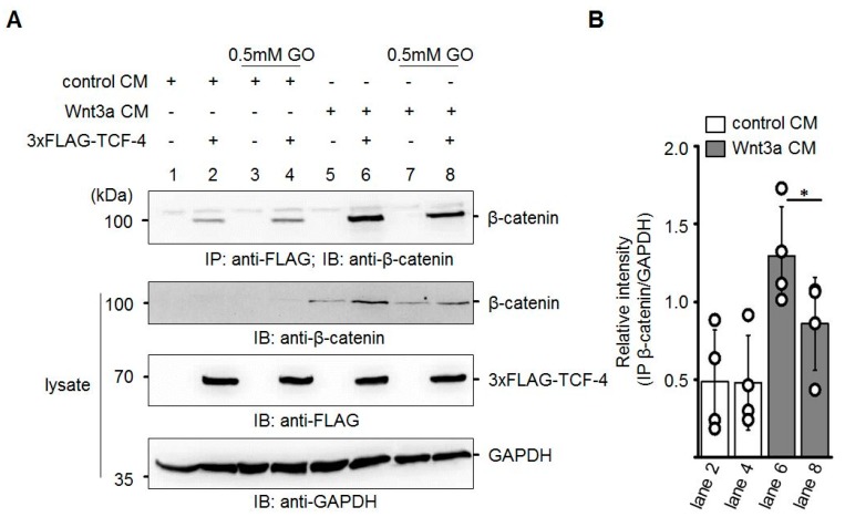
Figure 6.
Reduced binding of β-catenin to TCF-4 after GO-treatment of Wnt3a CM. (A) Neuro-2a cells were transiently transfected with 3xFLAG-TCF-4 and subsequently treated with the indicated CM. After immunoprecipitation (IP) with anti-FLAG antibody, the amount of co-precipitating β-catenin was analyzed by immunoblotting (IB). The presented blot is a representative of n = 4 independent experiments. (B) Quantification of Western blot experiments. Signal of co-immunoprecipitated β-catenin was normalized to GAPDH in the lysates. The graph shows mean values ±SD. Circles represent individual experiments. Data were analyzed with a paired t-test. * p < 0.05.

