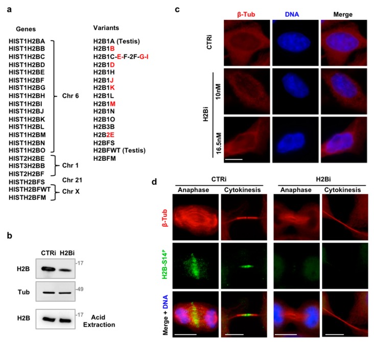
Figure 1.
Depletion of ecH2B with siRNAs. (a) Schematic representation of H2B isoforms (gene symbol, chromosomal location, and protein name are reported). In red are indicated the isoforms recognized by the nine variant-specific siRNAs. (b–d) HeLa cells were depleted for histone H2B with a combination of the nine variant-specific siRNAs (H2Bi) or with universal negative control (CTRi). Representative WB for the indicated proteins is shown. Cells were subdivided in two aliquots and cell extracts obtained with lysis buffer and centrifugation to eliminate chromatin or with acid extraction to purify nucleosome histones. (c) Representative IF imagines of CTRi and H2Bi HeLa cells—transfected with the indicated molarity of siRNAs—stained with Hoechst (blue) and anti-β-Tubulin Ab (red) to visualize nuclear DNA and cytoplasm. Scale bar is 10 μm. (d) Representative IF imagines of CTRi and H2Bi HeLa cells stained with anti-β-Tubulin Ab (red), anti-phospho-H2B-Ser14 Ab (green) and Hoechst (blue) to visualize nuclei. Representative images of cells in anaphase and cytokinesis are shown. At least 30 anaphase and 100 cytokinetic cells form three independent experiments were analyzed. Scale bar is 10 μm.

