Figure 2.
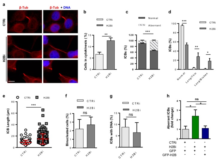
ecH2B depletion in HeLa cells induces cytokinesis defects. Proliferating, asynchronous HeLa cells were depleted for histone H2B, stained for β-Tubulin and DNA as described in Figure 1, and analyzed in panels A to F. (a) Representative IF images of cytokinetic cells. Compared to normal cytokinesis in CTRi (upper panels), the presence of long and thin (middle panels) or long and broken (lower panels) ICBs are shown in the H2Bi cells. Scale bar is 10 μm. (b) The percentage of cells in cytokinesis was measured by scoring at least 1000 cells per IF sample in four independent experiments. (c,d) The relative amount of normal and aberrant ICB was measured by scoring at least 1000 cells per sample and pooling (c) or subdividing (d) cytokinetic cells based on the defect type. (e) The length of each ICB was measured in three independent experiments. (f) The presence of cells with two or more nuclei and (g) ICBs with DNA (i.e., chromosome bridges, lagging chromosomes) was measured by scoring the same samples described in (b). (h) H2Bi and CTRi HeLa cells were transfected with GFP-H2B or GFP-empty vector and stained for β-Tubulin and DNA as above. The amount of long, aberrant ICB was measured by scoring at least 100 GFP-positive cells per sample. Data are reported as mean ± SD. ns p > 0.05; * p < 0.05; ** p < 0.01; *** p < 0.001.

