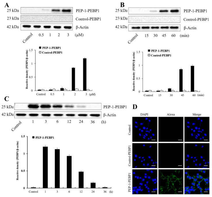
Figure 2.
Transduction efficacy and stability of control-PEBP1 and PEP-1-PEBP1 fusion protein in NSC34 cells. (A) Western blot analysis showing concentration-dependent (0.5–3 μM) cellular expression of polyhistidine for 1 h after PEP-1-PEBP1 and control-PEBP1 treatment. (B) Western blot analysis showing time-dependent (0–60 min) cellular expression of polyhistidine, analyzed after 3 μM PEP-1-PEBP1 and control-PEBP1 treatment. (C) Western blot analysis showing chronological (1–36 h) intracellular stability of polyhistidine expression for 1 h after PEP-1-PEBP1 treatment. (D) Immunocytochemical staining for polyhistidine in PEP-1-PEBP1 and control-PEBP1 protein for 1 h after PEP-1-PEBP1 treatment. Scale bar = 20 μm. The bars indicate mean ± SEM.

