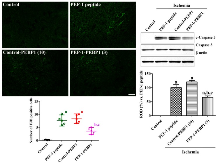
Figure 5.
Effect of Control-PEBP1 and PEP-1-PEBP1 protein on cell death and caspase 3 expression in the rabbit spinal cord against ischemic damage. Fluoro-Jade B (FJB) staining is conducted to detect the degenerating cells in the spinal cord 72 h after reperfusion. The number of FJB-positive cells in all the groups is also shown. Western blot analysis for caspase 3 and cleaved caspase 3 (c-caspase 3) in the spinal cord homogenates is conducted to compare the caspase 3 dependent neuronal death in spinal cord (n = 5 per group; a p < 0.05, significantly different from the control group, b p < 0.05, significantly different from the PEP-1 peptide-treated group; c p < 0.05, significantly different from the control-PEBP1-treated group). The bars indicate confidence interval or standard errors of mean.

