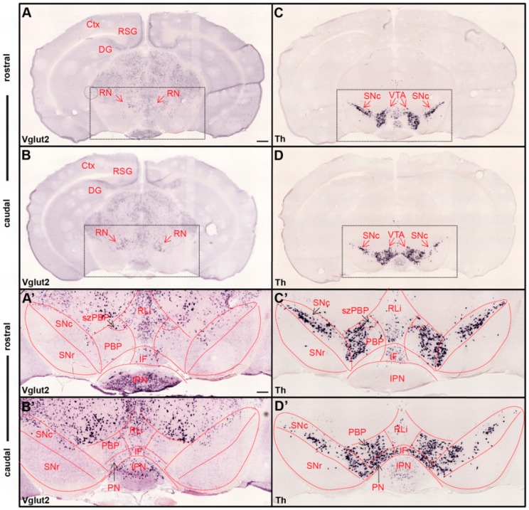
Figure 1.
Ample Vglut2 mRNA-positive cells throughout dorsal and ventral midbrain with more sparse expression within the dopaminergic area. Colorimetric in-situ hybridization showing overview of Vglut2 (A,B) and Th (C,D) mRNA in midbrain sections of wildtype adult mouse at two rostro-caudal levels. (A,B) Vglut2 mRNA is abundant throughout the midbrain with strong signals in e.g., the red nucleus (RN), retrosplenial group of the cortex (RSG) and dentate gyrus (DG), and weaker signals in the ventral tegmental area (VTA) and substantia nigra pars compacta (SNc). (C,D) Th mRNA is selectively localized in dopaminergic neurons of the VTA and SNc; its mRNA signal is implemented to visualize these areas. Dotted square around the VTA and SNc (scale bar 500 mm) presented as closeups in (A’–D’; scale bar 200 mm). (C’,D’) SNc, substantia nigra pars reticulata (SNr) and subregions of VTA outlined in Th closeups and superimposed on Vglut2 closeups (A’,B’). (C’,D’) Th mRNA was strongly localized in the SNc and within the parabrachial pigmented area (PBP) and paranigral nucleus (PN) of the VTA with a weaker signal in the rostral linear nucles (RLi) and caudal aspect of the interfascicular nucleus (IF). (A’,B’) Within the VTA, Vglut2 mRNA was detected in the PBP, PN, RLi, and IF as well as within the medially-located subzone of the PBP (szPBP) while no Vglut2 mRNA was detected in the GABAergic SNr area. Additional abbreviations: Ctx, Cortex; IPN, interpeducular nucleus. Reprinted from Papathanou et al., 2018 [24].

