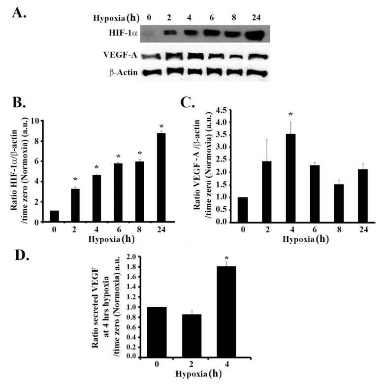
Figure 1.
Hypoxia mimicking increases the expression of hypoxia-inducible factor (HIF-1a) and the expression and secretion of vascular endothelial growth factor (VEGF) in glioblastoma cells. (A) SF-268 cells subjected to hypoxia using cobalt(II) chloride hexahydrate (CoCl2) up to 24 h. The total cell lysates were collected at different time intervals (2, 4, 6, 8, and 24 h post hypoxia or normoxia), as indicated, and the samples were blotted against HIF-1α, VEGF-A, or β-actin antibodies. Quantitation of HIF-1α (B) or VEGF-A (C) using ImageJ. The bands were normalized to β- actin and expressed as fold change compared to the control (normoxia). (D) SF-268 cells were subjected to hypoxia using cobalt(II) chloride hexahydrate (CoCl2) for 2 or 4 h or left in normoxic conditions. The supernatant was collected and measured for VEGF-A secretion (compared to standards) according to the manufacturer’s guidelines (described in Section 2). The data are means ± standard error of the mean (SEM) from three different experiments (n = 3); * p < 0.05 indicates statistically significant differences.

