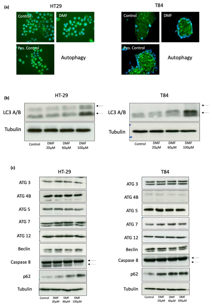
Figure 6.
DMF induces autophagy in HT-29 and T84 colon carcinoma cell lines. (a) representative immunofluorosecence of autophagy using Cyto-ID® assay. Cells were treated with DMF (100 µM) or solvent for 24 h. As a positive control Rapamycin (500 nM) + Chloroquine (10 µM) were used; (b) representative Western blot analyzes of LC3 A/B. Cells were treated for 24 h with vehicle or DMF for the indicated concentrations. The two arrows mark the type I (upper arrow) and type II (lower arrow) LC3A/B; (c) representative Western blot analysis from different autophagy associated proteins. Cells were treated for 24 h with vehicle or DMF for the indicated concentrations. Comparable results were obtained from at least three independent experiments.

