Figure 2.
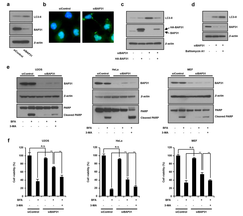
The suppression of BAP31 expression induces autophagy and antagonizes ER stress-induced cell death. (a) Loss of BAP31 increases LC3-II expression. U2OS cells were transfected with 150 pmol of siBAP31 or siControl for 24 h. Cells were subjected to immunoblotting using anti-BAP31, anti-LC3, and anti-β-actin antibodies. (b) U2OS cells stably expressing GFP-LC3 were transfected with 150 pmol of siBAP31 or siControl for 24 h. Cells were fixed with 4% PFA, and GFP-LC3 (green) fluorescence was determined. Blue represents nuclear DAPI staining. Scale bar, 10 μm. (c) U2OS cells were transfected with siBAP31 (+) or siControl (−) for 18 h and then transfected with HA-BAP31 (+) or pcDNA3.1 (−) for 12 h. Cells were subjected to immunoblotting using indicated antibodies. (d) BAP31 knockdown stimulates autophagosome synthesis. U2OS cells were transfected with 150 pmol of siBAP31 or siControl for 24 h, followed by treatment with or without 1 µg/mL of bafilomycin A1 for 1 h. Cells were subjected to immunoblotting using the indicated antibodies. (e,f) The suppression of BAP31 inhibits ER stress-mediated cell death by inducting autophagy. The cells were transfected with siBAP31 or siControl for 16 h and these cells were preincubated with or without 5 mM of 3-MA for 1 h and further incubated with or without BFA (1 µg/mL) for 18 h in U2OS, HeLa, and MEF cells. Cells were subjected to immunoblotting using the indicated antibodies (e) or MTT using cell viability assay (f). These experiments were repeated two times (a–e). Data are presented as the mean ± standard deviation (SD) of the three simultaneously performed experiments (f). P values were calculated using two-way ANOVA; n.s., not significant; ** P < 0.01 (f).

