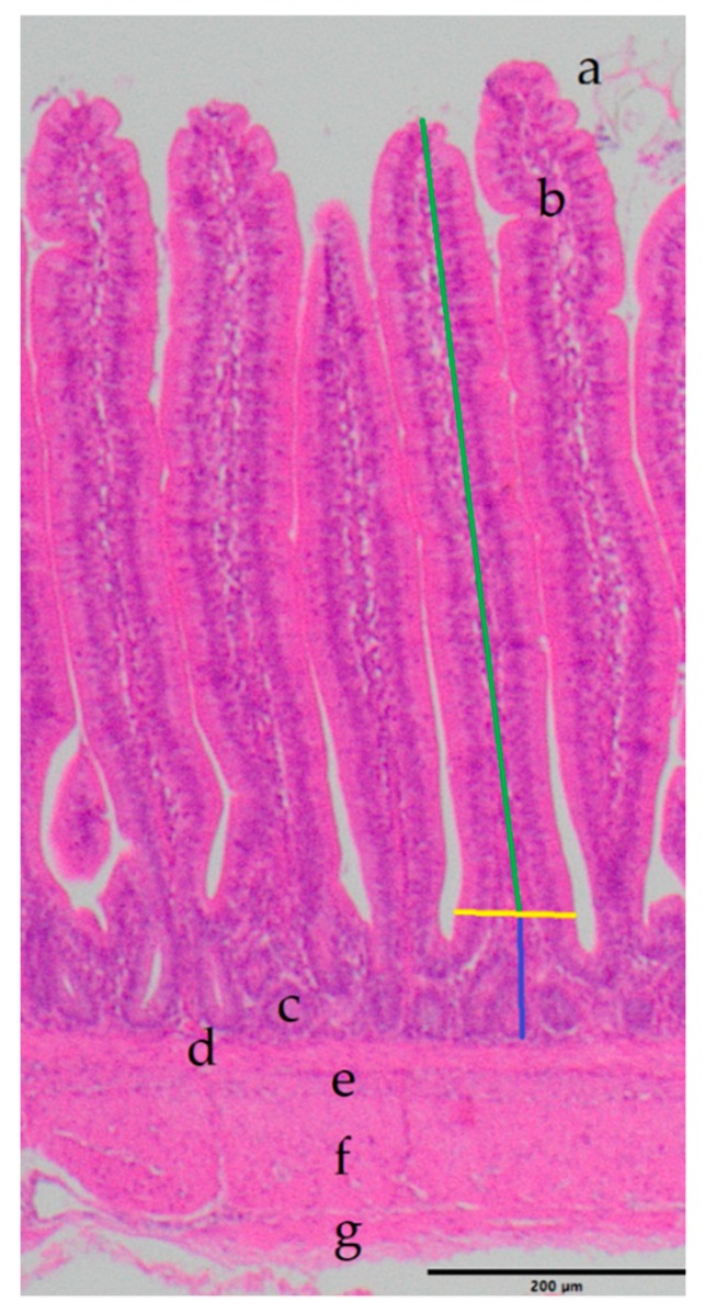
Figure 1.
Example image: HE staining from the ileal intestinal wall, measuring lines included. Scale bar = 200 µm. Green: height; yellow: width; blue: crypt depth; a = Villus intestinalis, b = Lamina propria mucosae, c = Lieberkuhn crypt, d = Lamina muscularis mucosae, e = Tela submucosa, f = Tunica muscularis, g = Tunica serosa.

