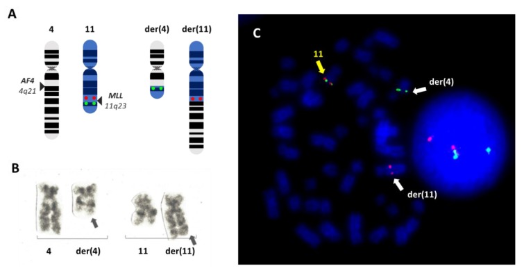
Figure 2.
The t(4;11)(q21;q23) rearrangement involving MLL. Schematic representation of the translocation between chromosomes 4 and 11 giving rise to the two derivative chromosomes der(4) and der(11) (A). The location of the fluorescence in situ hybridization (FISH) probe XL MLL (Metasystems) is also indicated on the normal chromosome 11, consisting of one green and one red signal flanking the MLL locus at 11q23. In the event of the translocation, the two signals split, indicating the disruption of the MLL locus. As a result, the der(11) retains the red signal proximal to MLL, while the der(4) will contain the green signal corresponding to the distal portion of MLL. These patterns are visible in a representative metaphase from the RS4;11 cell line, known to harbor a t(4;11)(q23;q21), hybridized with the XL MLL probe (B). The same rearrangement is shown in (C), obtained by G-banding.

