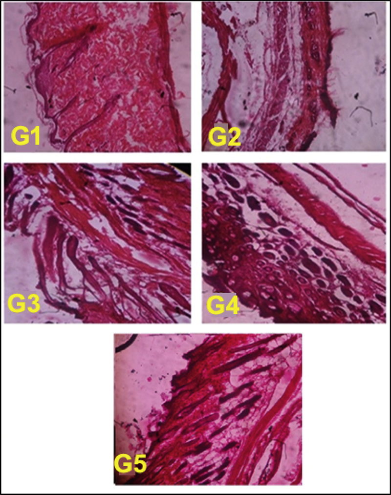
Fig. 8.
Melanoma affected skin histopathology of control (G1-G2) and treatment mice marketed and FCNGL formulation (G3-G5) are shown. The tissue architecture of non-tumor bearing mouse (G1) is well aligned; G2-mouse skin is completely broken with rupture of dermis layer; G3-mouse skin shows some degree of improvement in skin texture; in G4 and G5 treatment mouse, skin histology shows significantly healing with regeneration of keratinized squamous tissue. The tissue was visualized at 100X magnification using light microscope (Olympus, Japan), p < 0.05, n = 6.

