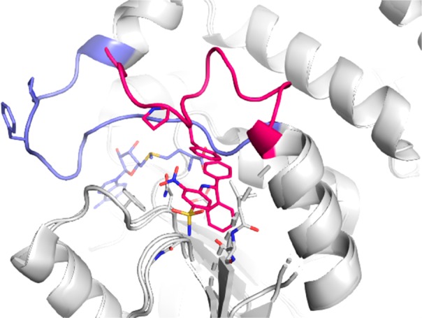
Figure 1.

Overlay of X-ray crystal structures of Dot1L with SAM (pdb 1nw3) and 1 (undisclosed structure). Cartoon representation of Dot1L (gray) with the flexible loop 126–140 shown in light blue for SAM and red for 1; stick models of ligands 1 (red) and SAM (blue). The residues Phe129 and Pro130 that form the roof of the induced pocket are shown as sticks.
