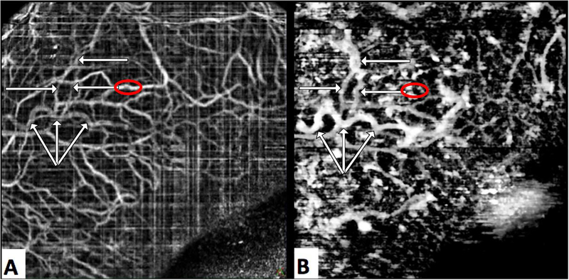
Figure 5.
Frames A and B were derived from a single OCT-A scan. Frame A is the conventional OCT-A image comprised of a map of moving reflective sources presumed to be blood cells. Frame B is a is a map of scleral voids; presumed to be areas in which clear aqueous create dark openings in OCT structural scans. Aqueous humor outflow vasculature (white arrows) produce a strong “signal” map of scleral voids (B), but are faintly visible in the OCT-A image (A) suggesting that the aqueous within contains some scattering media. Prominent blood vessels (red circle, A) present as shadows in the map of scleral voids (red circle, B).

