Fig. 3.
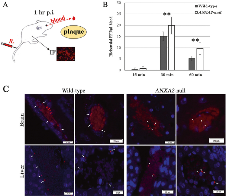
Global depletion of ANXA2 blocked rickettsial adherence to the blood vessel luminal surface in vivo. a The anatomically based in vivo quantitative rickettsial (R.) adhesion analysis system. b Plaque assay for R. australis using blood samples collected from the orbital venous sinus of WT (n = 10) and ANXA2-null (n = 10) mice at different time p. i. with 10 LD50 doses of R. australis intravenously. Data are represented as mean ± SEM. **Compared to the control group, P < 0.01. c Representative IF-based identification of rickettsial (red) location in WT and Anxa2-null mice 60 min p.i. with 10 LD50 doses of R. australis intravenously. Rickettsiae are adhered to intima layer of blood vessels (arrows) or wrapped in blood clots (arrowheads) in the lumens of blood vessels. Nuclei are blue. Scale bars; 20 μm

