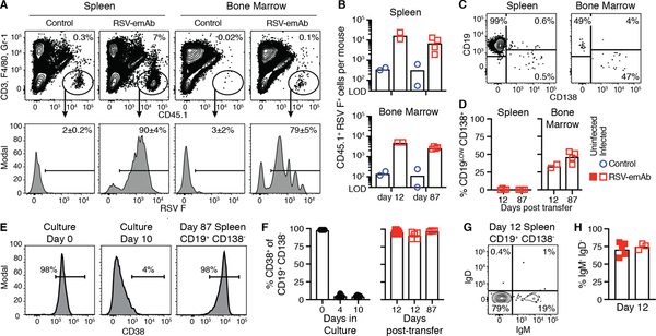
Figure 8. Phenotype of RSV-emAb B cells in immunocompromised mice.
(A) Representative flow cytometric analysis and (B) quantitation of CD45.1+ RSV F+ CD3− F4/80− Gr-1− Fixable viability dye (FVD)− donor B cells in spleen and bone marrow from individual RAG1−/− mice from two combined experiments 12 or 87 days after transfer of 1.5 × 10⁷ control or RSV-emAb B cells (n=2–4). Samples were enriched for CD45.1+ cells prior to analysis and all mice were infected with RSV five days prior to analysis. The limit of detection (LOD) was established in samples from mice that did not receive cell transfer (n=7). (C) Representative flow cytometric analysis and (D) quantitation of the % of CD19LOW CD138+ RSV-emAb B cells in the spleen and bone marrow from individual mice (n=2–4) from two combined experiments. (E) Representative flow cytometric analysis and (F) quantitation of CD38 expression by CD19+ CD138− RSV-emAb B cells in the spleen of recipient mice compared to B cells at start of culture and at the time of cell transfer (n=2–5) from three combined experiments. (G) Representative flow cytometric analysis and (H) quantitation of % of CD19+ CD138− RSV-emAb B cells in the spleen of individual animals that were IgM− IgD− twelve days after transfer (n=3–5).

