
Figure 5.
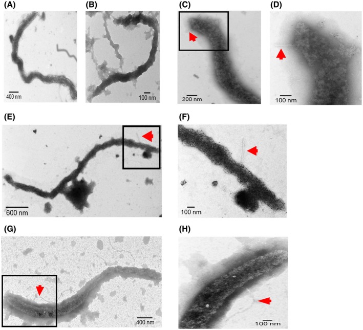
Transmission electron microscopy analysis of ‘Candidatus Liberibacter asiaticus’ (Las). (A) Las cell isolated from Las‐infected grapefruit seed coat. (B) Las cell isolated from dodder stem. (C)–(H) Las isolated from psyllid guts. (D), (F) and (H) are enlarged views of the indicated area in (C), (E) and (G), respectively. Las cells were visualized by using negative staining and antibody against Las OmpA and second antibody anti‐rabbit IgG‐gold. Arrows indicate thread‐like structures. Size bars are indicated in the images.