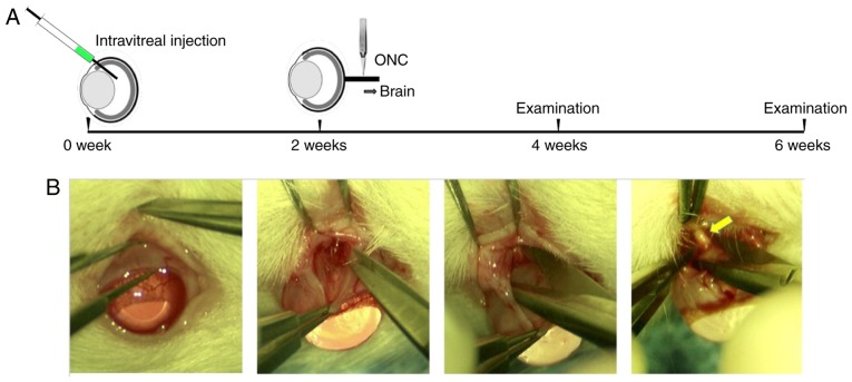
Figure 1.
Experimental design. (A) Schematic timeline of the experimental procedure. Two weeks after intravitreal injection of adeno-associated virus vectors, ONC was performed on the injected eyes. Analysis, including morphological, functional and electrophysiological changes of RGCs/ONs, was performed prior to ONC, and 2 and 4 weeks after ONC. (B) ONC procedure (magnification, ×6). The yellow arrow indicates the site of the ONC. ONC, optic nerve crush; RGC, retinal ganglion cell.

