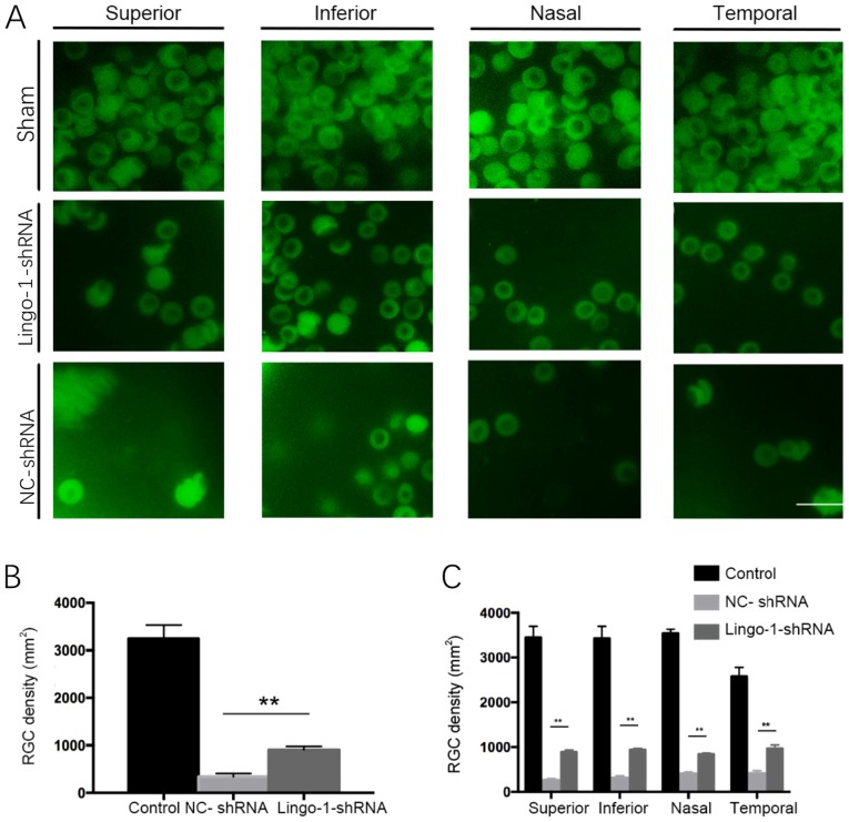
Figure 3.
RGC survival in different groups following optic nerve crush injury. Two weeks after optic nerve crush, surviving RGCs were observed in the in the superior, inferior, nasal and temporal quadrants of retinal flat mounts via immunofluorescent staining for RNA-binding protein with multiple splicing. Representative images were captured at 1 mm from the optic disc. (A) Representative images of RGCs treated with sham control, NC-shRNA or AAV2-lingo-1-shRNA following optic nerve injury. Scale bar=50 µm; magnification, ×100. (B) Quantitative analysis of the average number of surviving RGCs in whole retina samples. (C) Density of surviving RGCs in different retinal quadrants. **P<0.01. Lingo-1, leucine-rich repeat and immunoglobulin-like domain-containing nogo receptor-interacting protein 1; RGC, retinal ganglion cell; NC-shRNA, negative control shRNA; shRNA, short hairpin RNA.

