Figure 5.
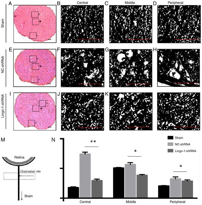
Transverse histological sections of optic nerves. (A) Representative histological section of the sham ONC group. Details of histological alterations of the (B) central, (C) middle and (D) peripheral part of the optic nerve of the sham ONC group. (E) Representative histological section of the NC-shRNA group. Details of histological alterations of the (F) central, (G) middle and (H) peripheral part of the optic nerve of the NC-shRNA group. (I) Representative histological section of the lingo-1-shRNA group. Details of histological alterations of the (J) central, (K) middle and (L) peripheral part of the optic nerve of the lingo-1-shRNA group. ★, central area; ☆, middle area; °, peripheral area. Scale bar=100 µm. (M) Schematic drawing showing the observation site of transverse histological sections of optic nerves. (N) Quantitative analysis of nerve cavity areas in the three groups revealed that the injury damaged the central, middle and the peripheral parts of optic nerves, and the damage was most severe in the central parts of the optic nerves. n=5. *P<0.05 and **P<0.01. Lingo-1, leucine-rich repeat and immunoglobulin-like domain-containing nogo receptor-interacting protein 1; ONC, optic nerve crush; NC-shRNA, negative control shRNA; shRNA, short hairpin RNA.

