Figure 6.
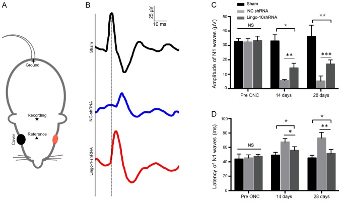
Evaluation of the recovery of injured optic nerves using the F-VEP wave pattern. (A) Locations of silver needle electrodes. ▲, the reference electrode; ★, the recording electrode; ✦, the ground electrode. The red color indicated the testing eye of the rat. (B) Representative F-VEP tracings 4 weeks following ONC in the sham surgery, NC-shRNA and lingo-1-shRNA groups. Y-axis scale, 25 µV; x-axis scale, 10 ms. (C) N1 amplitude 2 and 4 weeks following ONC. (D) N1 latency 2 and 4 weeks following ONC. Error bars represent standard error of the mean, n=10. *P<0.05, **P<0.01 and ***P<0.001. Lingo-1, leucine-rich repeat and immunoglobulin-like domain-containing nogo receptor-interacting protein 1; NC-shRNA, negative control shRNA; shRNA, short hairpin RNA; F-VEP, flash visual evoked potential; NS, not significant.

