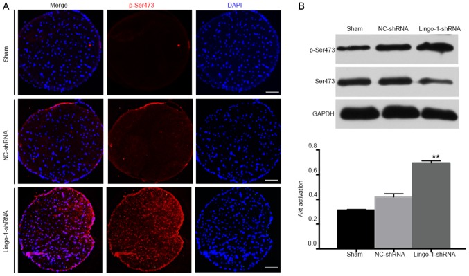
Figure 7.
p-Ser473 expression in optic nerves. (A) Representative immunofluorescent staining images presenting p-Ser473 expression (red; scale bar=100 µm) in sham surgery, NC-shRNA and lingo-1-shRNA groups. (B) Western blot analysis of Akt activation of the injured optic nerves. The ratio of p-Akt to total Akt expression levels was calculated four weeks following optic nerve injury. Total protein level was normalized to β-actin levels, n=5. **P<0.01 vs. the NC-shRNA group. Lingo-1, leucine-rich repeat and immunoglobulin-like domain-containing nogo receptor-interacting protein 1; p-, phosphorylated; NC-shRNA, negative control shRNA; shRNA, short hairpin RNA; Akt, protein kinase B.

