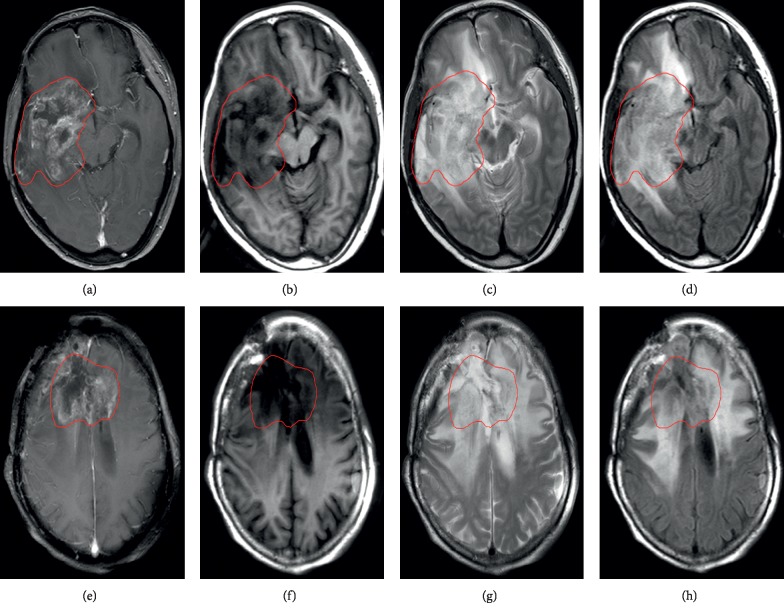
Figure 1.
MRI diagnostic images of two patients with glioma. (a–d) Recurrent image of a 52-year-old patient with astrocytoma 1 year after radiotherapy. (e–h) Necrotic image of a 54-year-old patient with oligodendroglioma 6 months after radiotherapy. (a), (b), (c), and (d) and (e), (f), (g), and (h), respectively, show T1C, T1, T2, and FLAIR images. The inside of the red line shows the edge of the lesion.

