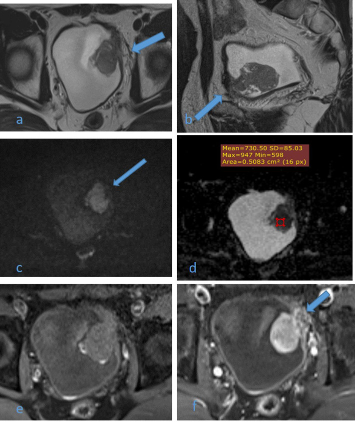
Figure 4.
A 54-year-old male patient with history of recurrent hematuria. High resolution axial (a) and sagittal (b) T2WI images shows muscle-invasive left anterolateral U.B. polypoidal lesion disrupting the underneath hypointense detrusor lining, with no extension to perivesical fat (arrows). DW image (c) and ADC map image (d) show highly restricted diffusion of mass with facilitated diffusion of muscle layer exclude muscle invasion (arrow) and low ADC value about 0.7 × 10⁻³ s/mm2 denoting malignant nature, DCE-MRI (e) non-contrast subtracted image and (f) early arterial phase image are displaying early intense enhancement of the mass with interrupted enhancement of muscle layer denoting muscle invasion (arrow). Multiparameteric MRI are consistent with muscle-invasive bladder cancer (Stage T2) and VI-RADS IV which confirmed histopathologically. ADC, apparent diffusioncoefficient; T2WI,T2 weightedimaging; VI-RADS, vesical imaging reporting and data system.

