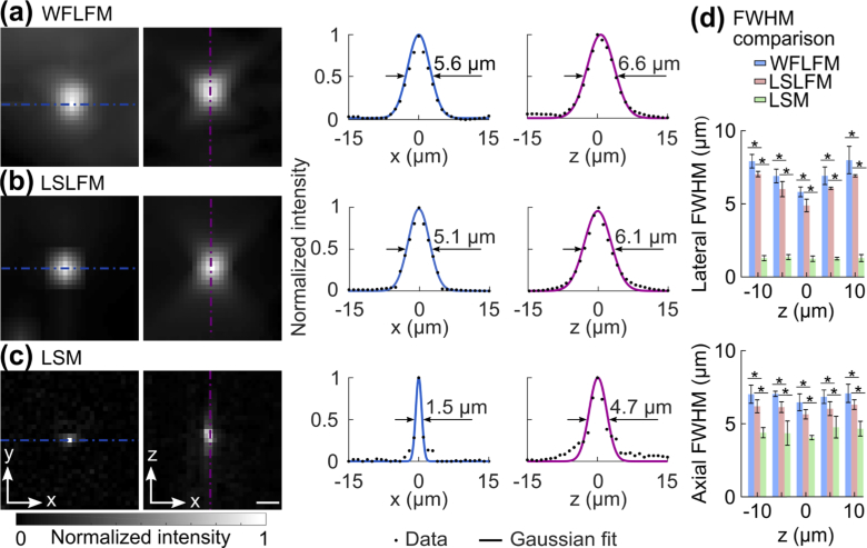
Fig. 4.
Imaging of sub-diffraction beads demonstrated that LSLFM imaged with finer resolution than WFLFM. Images of individual 0.5 µm beads and the intensity profiles along the dashed lines obtained with (a) WFLFM, (b) LSLFM, and (c) LSM. Fits to Gaussian profiles determined the FWHM. Scale bar: 5 µm. (d) Comparison of FWHM between the three tested imaging modalities (mean ± std.). The lateral and axial FWHMs for LSLFM were smaller than those of the WFLFM, *p < 0.05 (n = 6, two-sided Wilcoxon rank-sum test).

