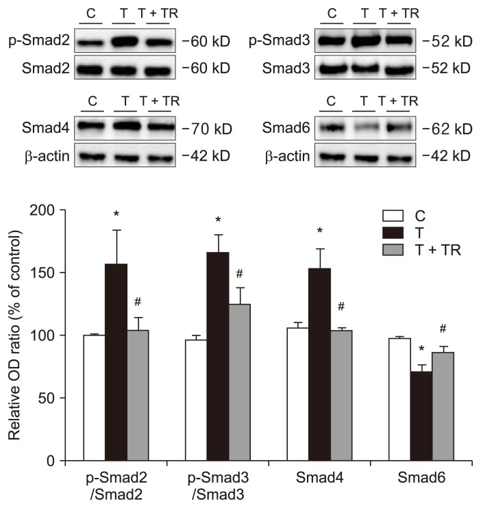
Figure 3.
Effects of tranilast on the Smad-dependent signaling pathway.
Western blot analysis was performed on human peritoneal mesothelial cells (HPMCs) exposed to transforming growth factor-beta 1 (TGF-β1) in the presence or absence of tranilast (100 μM) for 24 hours. Quantification is relative to the control and normalized to β-actin expression. TGF-β1 increased the p-Smad2/Smad2 and p-Smad3/Smad3 protein expression ratios, increased Smad4 protein expression, and reduced Smad6 protein expression. Tranilast reversed these changes. n = 4 per group.
C, control; OD, optical density; T, HPMCs with TGF-β1 treatment; T + TR, HPMCs with TGF-β1 and tranilast cotreatment.
*P < 0.05 compared to the C group, #P < 0.05 compared to the T group.

